Fine Needle Aspirate (FNA) for Dogs, Cats, and Other Pets
TL;DR (Too Long; Didn’t Read)
A Fine Needle Aspirate (FNA) is a quick, minimally invasive way for veterinarians to collect cells from lumps, masses, or fluid accumulations for microscopic examination. It helps determine if a growth is benign, malignant, or inflammatory. While almost all veterinary clinics can collect FNA samples, not all can process them in-house—some send them to specialized labs for review by a veterinary pathologist. Southern Ocean Animal Hospital has the advanced tools, labs, and surgical expertise to both collect and interpret FNA results quickly, often during the same visit.
ELI5 (Explain Like I’m 5)
Imagine your pet has a little bump. Instead of doing surgery right away, the vet uses a tiny straw-like needle to take just a few cells from that bump—kind of like sipping a bit of a milkshake. Then, they look at those cells under a special “super zoom” camera (microscope) to see if the bump is harmless or something that needs more attention.
Summary of Key Points
- What It Is: FNA uses a small needle to collect cells from lumps, masses, internal organs, or fluid.
- Why It’s Done: Helps identify infections, inflammation, or cancer without invasive surgery.
- Equipment: Needle, syringe, microscope slides, stains, alcohol swabs—plus advanced imaging (if guided).
- In-house vs. External: Many clinics collect samples but send them to outside labs for analysis. Southern Ocean Animal Hospital has on-site diagnostic equipment for rapid results.
- Benefits: Quick, low-cost, low-risk way to get crucial diagnostic information.
- Limitations: Not always conclusive—sometimes requires follow-up biopsies.
- Timeline: Can range from minutes (in-house) to a week (external lab) for results.
- Scenario: Pet owner brings in dog with a fast-growing lump—FNA done in minutes, results same day, treatment started right away.
- Systems: Works for dogs, cats, and many exotic pets.
What is a Fine Needle Aspirate?
A Fine Needle Aspirate (FNA) is a veterinary diagnostic procedure used to collect a small sample of cells from a suspicious area—such as a lump, swollen lymph node, fluid pocket, or internal organ—so they can be examined under a microscope. This method provides valuable insight without the need for surgical biopsies, making it safer, faster, and less stressful for pets.
Why FNAs Are Used
Veterinarians recommend FNAs when:
- New lumps appear on your pet’s skin or under the skin.
- Existing lumps change in size, shape, or texture.
- Organs look abnormal on X-ray, ultrasound, or physical exam.
- There’s swelling in lymph nodes or other tissues.
- Fluids accumulate in the chest, abdomen, or joints.
FNAs help answer crucial questions:
- Is the lump benign or cancerous?
- Is the swelling due to infection, inflammation, or a tumor?
- Does this require immediate surgery, medical management, or no treatment?
Do All Vet Clinics Have FNA Equipment?
Basic Capability
- Most veterinary clinics have the basic tools to perform an FNA:
- Sterile needles (usually 22–25 gauge)
- Syringes (3–12 mL)
- Microscope slides
- Alcohol wipes
- Fixative solutions (like methanol or specific cytology fixatives)
- These tools are inexpensive and widely available, so FNA collection itself is almost universally possible.
In-house vs. External Processing
- In-house labs: Clinics with microscopes and trained staff can process and interpret the sample during the visit.
- External labs: Samples can be sent to specialized pathology labs, where board-certified veterinary pathologists examine them.
Southern Ocean Animal Hospital Advantage
- Has on-site lab equipment, including microscopes, advanced staining systems, and imaging tools.
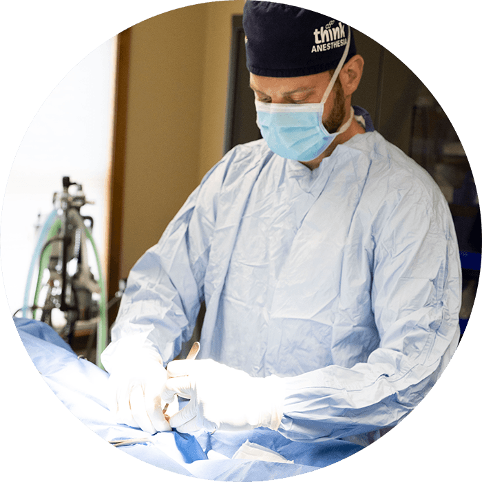
- Under the guidance of Daniel N. Pascetta, DVM, our skilled veterinary team is experienced in performing Fine Needle Aspirates (FNA) on-site and delivering prompt, accurate results. Dr. Pascetta’s leadership ensures that every case benefits from his extensive clinical expertise, allowing our team to assess samples quickly and create effective treatment plans tailored to each pet’s needs.
- Ultrasound-guided FNAs are available for sampling internal organs safely.
FNA Procedure: Step-by-Step
1. Physical Examination
The veterinarian feels the lump or examines imaging to determine the best sampling spot.
2. Preparation
- The site is cleaned with alcohol or an antiseptic.
- Sedation is rarely needed unless the pet is anxious or the lump is in a sensitive location.
3. Needle Insertion
- A fine needle attached to a syringe is inserted into the mass.
- Gentle suction is applied to collect cells.
4. Sample Transfer
- Cells are expelled onto microscope slides.
- The sample is either air-dried or fixed with a spray fixative.
5. Staining & Microscopy
- If processed in-house, slides are stained with special dyes (e.g., Diff-Quik) for cellular detail.
- A trained veterinarian or technician examines the sample.
6. Sending to a Pathologist
- For more complex cases, slides are sent to a diagnostic lab.
Uses of FNA in Veterinary Medicine
- Skin and Subcutaneous Lumps
- Lipomas (benign fatty tumors)
- Mast cell tumors
- Sebaceous cysts
- Abscesses
- Internal Organs
- Liver
- Spleen
- Lymph nodes
- Kidneys
- Fluid Sampling
- Joint fluid for arthritis or infection
- Abdominal or chest fluid for disease detection
Benefits of Fine Needle Aspirates
- Quick: Many results available within the same visit.
- Minimally Invasive: No incisions, stitches, or prolonged recovery.
- Low Cost: Less expensive than surgical biopsies.
- Versatile: Can be used for many types of tissues and fluids.
Limitations
- Non-diagnostic samples: Sometimes the needle collects too few cells.
- Sample bias: The needle may miss abnormal areas.
- Need for confirmation: Suspicious results often require follow-up biopsies.
Example Timeline
- Day 1: Pet seen for new lump → FNA performed → preliminary in-house results given.
- Day 2–5: If sent to the lab, results returned from the veterinary pathologist.
- Day 5–7: Treatment plan finalized based on diagnosis.
Example Scenario for Pet Owners
Mrs. Lee brings her 9-year-old Labrador to Southern Ocean Animal Hospital after noticing a fast-growing lump on its side.
- Dr. Pascetta examines the lump and performs an FNA during the same appointment.
- Within 30 minutes, in-house cytology shows the lump is likely a benign lipoma.
- No surgery is needed, but the lump will be monitored.
- The quick diagnosis avoids unnecessary surgery, anesthesia, and cost.
Systems & Specialized Equipment
Southern Ocean Animal Hospital uses:
- High-quality microscopes for cytology.
- Diff-Quik and Wright-Giemsa staining systems.
- Ultrasound for guided FNAs of internal organs.
- Full in-house lab for rapid diagnostics.
FNA for Dogs, Cats, and Other Pets
While most FNAs are done on dogs and cats, they are also used for:
- Rabbits (abscesses, tumors)
- Ferrets (adrenal masses, lymphoma)
- Birds (skin nodules)
- Reptiles (masses, abscesses)
Southern Ocean Animal Hospital Location
Southern Ocean Animal Hospital is located at 319 E Main St, Tuckerton, NJ 08087, and proudly serves pets and their families across Ocean County and well beyond. Our service area includes nearby communities such as Stafford Township, Barnegat, Waretown, LBI, Long Beach Island, Beach Haven, Ship Bottom, Surf City, Long Beach Township, Tuckerton, Little Egg Harbor, and Eagleswood. We also regularly welcome clients from extended areas, including Lacey Township, Forked River, Bayville, Ocean Gate, Seaside Heights, Point Pleasant, Brick, and Toms River, as well as many parts of the Jersey Shore. Our reach extends into Atlantic County towns such as Galloway, Absecon, Brigantine, and Atlantic City, plus areas of Burlington County, including New Gretna and Bass River Township.
Our modern facility is fully equipped with advanced diagnostic and surgical tools, on-site laboratory capabilities, and specialized imaging technology. This allows us to perform Fine Needle Aspirates (FNA), interpret results quickly, and create effective treatment plans—often during the same visit—for the best possible outcomes for pets and peace of mind for their families.
Dr. Daniel N. Pascetta’s Professional Experience
Dr. Pascetta is a highly experienced veterinarian and surgeon with a strong diagnostic background. His approach to FNAs focuses on:
- Rapid diagnosis: Minimizing owner anxiety by providing same-day results when possible.
- Precision sampling: Using ultrasound-guided FNAs for internal masses.
- Comprehensive care: Combining FNA results with other diagnostics (bloodwork, imaging) for complete health assessments.
- Client communication: Ensuring owners understand the results, options, and next steps.